Racing To The End
Week seven of summer research is coming to a close and we are racing to the end, literally.
Last the whole lab participated in the B Well 5k on campus and sported our amazing new lab t-shirts designed by Khi Kim.
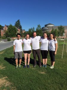
Williams Lab (L to R): Quang, Larissa, Derek, Maddie, and Alex
Going into the final week everyone is working to finish up the last parts of our projects. Alex is working through the ChiP protocol and doing some final tune ups. Hopefully next week she will be able to run through the whole procedure to see if there are any other places it could be improved. Quang is working away at getting through the data and making some big strides in figuring out the software needed to visualize his finding.
Derek and I have been learning how to use the scanning electron microscope (SEM) to visualize the neuromasts. To do this though we have had to enlist the help of some other amazing Carnegie Science faculty. SEM only allows you to see surface level detail so Nancy Kleckner and her extremely steady hands has been helping us dissect tissue off to expose the structures we want to image. The fish are only 96 hours old and only about 3.5mm long so we have to be extremely gentle and patient so we don’t ruin the tissue. Greg Anderson, the resident SEM guru, has been taking Derek and I through the preparation of the fish for imaging. They need to dried using a critical point dryer, which uses CO2 and heat to dry the tissue without damaging any of the cells. After drying the fish need to be coated in a thin layer of something metallic. This was my favorite part because we got to use the sputter coater which makes this purple plasma cloud.
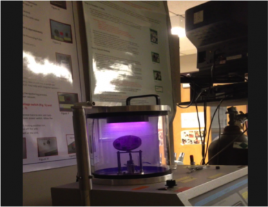
We turned off the lights for this part to get the full effect! Finally after all that prep, we could image our samples. We were able to get a few images of both lateral line neuromasts and the inner ear.
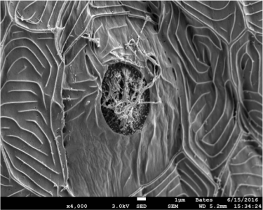
Lateral line neuromast
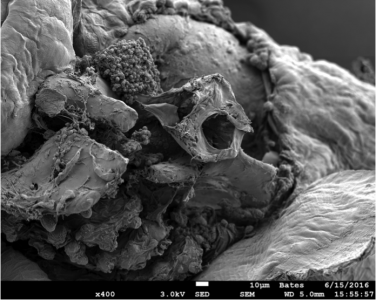
Otic vesicle with the otoliths removed. The circular bony structure is the semi-circular canal with some neuromasts surrounding the structure.
Next we are going to look at the effects of fish treated with oxidative stress inducing chemicals and see if we can see some structural differences with the SEM.