Forms of Resonance
In an accident at home in September 2007, longtime Bates staffer Sylvia Deschaine sustained head injuries that nearly killed her.
During her long recovery, she was fascinated by MRIs — magnetic resonance imaging scans — showing how the golf ball-sized opening in her skull, sustained falling down a flight of stairs, was healing.
When Deschaine came back to work, as the administrative assistant for the education and psychology departments, she seemed much the same as ever, dry wit and Irish brogue intact. But at heart she felt renewed, viewing others with a new compassion and concern, she says.
In that sense, she says, the accident is “probably one of the best things that has happened to me.”
Grateful for the hospital care she had received, Deschaine wanted somehow to bring the College and the local medical establishment closer together so as to support Bates students aiming for healthcare careers. This was part of her healing too, she says.
She discussed her experiences, especially the MRI, with Nancy Koven, assistant professor of psychology. A clinical neuropsychologist, Koven was already using MRI images of the brain in her research and teaching.
“Sylvia has a patient’s view, and I have a researcher’s view,” says Koven. “When we were in the hallway trying to have the same conversation, it meant different things to each of us.”
As they reconciled those perspectives, Koven realized that her students, too, could benefit from the patient’s view. “It’s a piece I just can’t do in the classroom,” she says. When Deschaine suggested a class visit to an MRI facility, it seemed like just the thing for one Koven course in particular.
The course, “Cognitive Neuroscience,” relates brain structure to function. Koven divides the coursework between anatomy and cognition. Teaching the former involves MRIs of healthy brains; the latter includes pencil-and-paper tests whose numerical scores indicate cognitive capabilities.
The two types of data complement each other for stronger diagnoses, Koven says. But a third kind of information helps, too — a patient’s presenting symptoms, medical history, lifestyle.
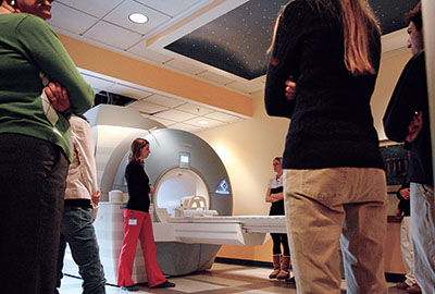
And so, one February day, the “Cognitive Neuroscience” class has just finished inspecting the new MRI scanner at St. Mary’s Regional Medical Center. The students are now viewing scans in a darkened room next door.
Barbara Mandy, a magnetic resonance imaging technologist, is showing the scans, which resemble X-rays. She pulls up a patient’s brain, scanned this morning. “Uh-oh,” she says. “This gentleman has no idea what’s growing in him.”
The patient — anonymous to the students — came to St. Mary’s complaining of memory and motor-skills problems. Koven asks her students to identify the pathological portion of the patient’s brain and suggest how a tumor, indicated by a dark patch in the image, might affect his mood or behavior — his life.
Which someone does. They’ve learned their lessons well, these strangers, seeing on a screen evidence of the most private kind of trouble for one scared man.
That’s a lesson Sylvia Deschaine gave to the class. “Just like that, we can be gone,” she says. “We’ve got to value each other.”