Microscopy
Bates supports a wide variety of microscopy techniques (see below). These instruments are available to all Bates faculty, staff, and students. For training or general consultation, please contact Mark Lessard.
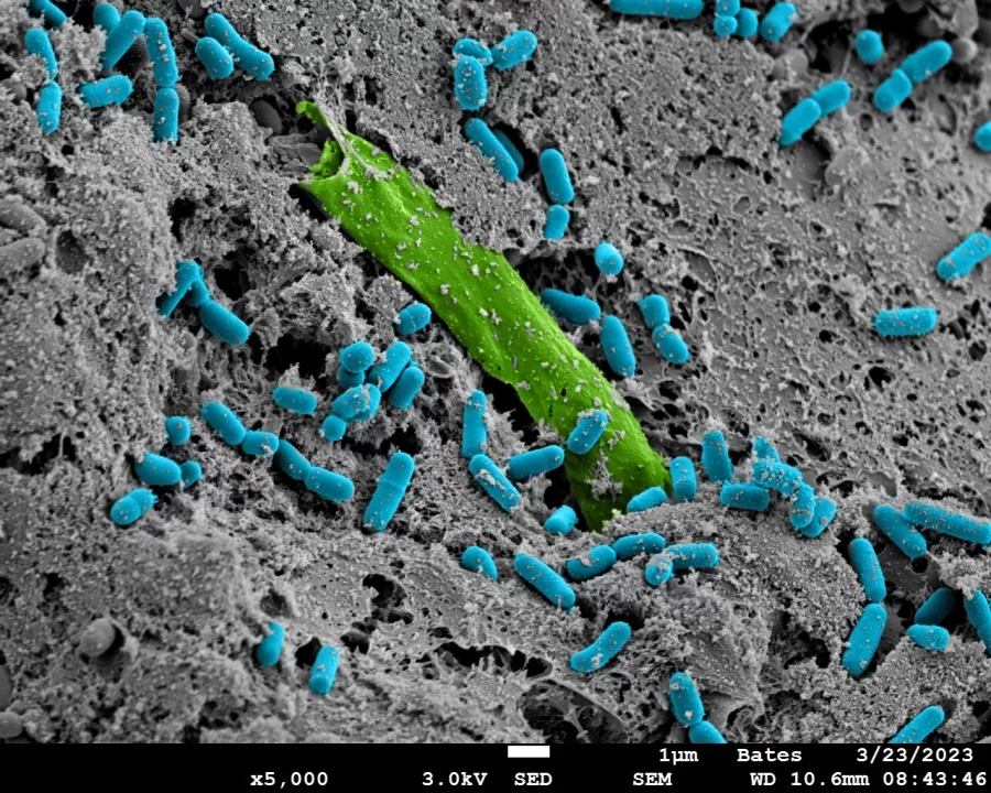
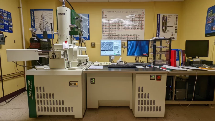
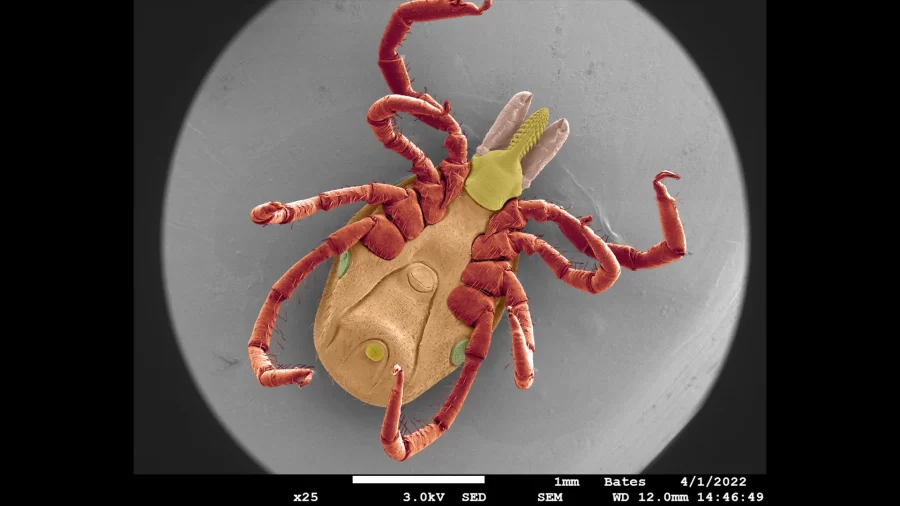
+JEOL JSM-7100FLV
Field Emission Variable Vacuum Scanning Electron Microscope with EDS
This instrument is equipped with a sample chamber-level secondary electron detector and both HV and LV backscatter detectors. It is equipped with a Schottkey thermal field emission gun and offers resolution of approximately 2-3 nm at 30 kV.
The SEM is equipped with a Thermo/ Noran System 7 Energy Dispersive Spectrometer with a LN-free digital detector. This system enables quantitative spectrum analysis with and without standards, spectral imaging, x-ray mapping, x-ray line scans, point and shoot sampling, and digital image acquisition.
Sample preparation equipment for the SEM:
Denton Vacuum DV1 Carbon Evaporation unit; attachments for rods or yarn
Anatech Hummer 6.6 Sputtering System with rotating, tilted stage and Au/Pd target
Ladd Research CO2 Critical Point Dryer
Scandium Image analysis software
Adobe Photoshop software
Fume hood
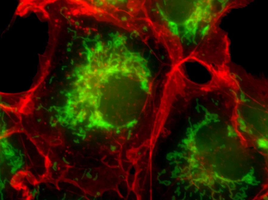
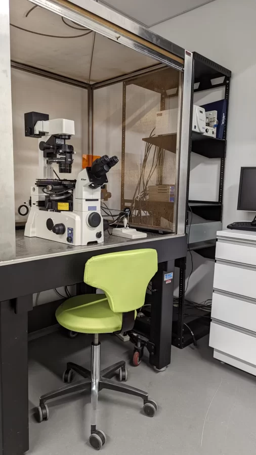
+Leica SP8 Laser Scanning Confocal Microscope
The Leica SP8 confocal microscope is equipped with the following:
- 405nm laser, Argon laser (458, 476, 488, 496, 514nm lines), White Light Laser (470nm -670nm), excitation sources
- 5 fluorescence detectors (2 PMT’s, 3 HyD’s), and a transmitted light detector
- Objectives:
- 10x 0.35NA
- 20x 0.75NA multi-immersion
- 25x 0.95NA water immersion
- 40x 1.1NA water immersion with motorized correction collar
- 40x 1.3NA oil immersion
- 63x 1.4NA oil immersion
- Full environmental chamber for live imaging
+Other light microscopes available:
Nikon Eclipse TE2000-U Widefield Fluorescence Scope (Inverted)
Configuration:
- Motorized stage, focus, and shutters
- Excite 120 fluorescence light source
- 5 filter cube positions
- Nikon Digital Sight DS-Qi1Mc Camera (monochrome, 1.5 megapixel, cooled)
- Software: ftp://freeware:freeware@ftp.nisoftware.net/
- https://www.nisoftware.net/NISoftware/Default.aspx
- Objectives:
- 4x 0.13 NA Plan Fluor
- 10x 0.3 NA ELWD Plan Fluor
- 20x 0.45 NA ELWD Plan Fluor
- 40x 0.6 NA ELWD Plan Fluor
Nikon Eclipse 80i Fluorescence Scope (Upright)
Configuration:
- Automated stage and focus
- 6 filter cube positions
- XCite 120 Fluorescent Source
- QImaging Retiga EXi Camera (mono and RGB capable)
- Objectives:
- 4x 0.2 NA Plan APO
- 10x 0.45 NA Plan APO
- 20x 0.7 NA Plan APO
- 40x 0.95 NA Plan APO
Nikon Modular Focus Scope (Upright)
- Configuration:
- Automated stage and focus
- Nikon Digital Sight DS-Ri1 Camera
- 100w Halogen BF source
- XCite 120 Fluorescence Source
- FiberLite Transillumination source
- FiberLite Gooseneck source
- Objectives:
- 2x 0.1 NA Plan APO
- 5x 0.15 NA LU Plan Fluor
- 10x 0.3 NA LU Plan Fluor
- 20x 0.4 NA ELWD LU Plan
- 50x 0.55 NA ELWD LU Plan
- Notes:
- This is a unique system that crosses a stand from a dissection scope with the imaging head of a standard upright scope.
- It is capable of fluorescence, brightfield, darkfield, DIC, Phase, and Polarization