Between Science and Art
X-ray Photographs by Judith K. McMillan
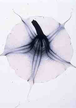
Judith K. McMillian Brugmansia x candida (Angel’s Trumpet), 2000 toned gelatin silver print, 16×20 inches Collection the artist, courtesy Cleveland Museum of Natural History
January 14 – March 19, 2005
Using an X-ray machine as her camera, contemporary Ohio artist Judith K. McMillan (American, born 1945) photographs the internal structures of plants, revealing the beauty of natural forms invisible to the human eye. Fascinated by natural cycles, the artist uses specimens of plant and insect material collected throughout the seasons.
Combining a documentary style with an eye for composition, McMillan’s images exist in a realm between science and art. Her unique depictions of nature have been described as simultaneously ephemeral and eerie. What are thought of as familiar forms, such as magnolia blooms or fern leaves, are transformed in her X-ray photographs in revealing and unexpected ways.
McMillan used an X-ray machine at The Cleveland Museum of Natural History to make these photographs. After a period of experimentation, she began creating photograms on film – life-sized depictions of the botanical material that she placed in the machine. Through methodical tests she determined the level of radiation that allowed the X-ray film to function as photographic negatives to be used to make the final photographs. McMillan then toned the prints to produce warm and cool values that add dimension and texture.
The artist chooses her subjects for their hidden forms, thereby rendering common plants and trees, such as dogwood branches or rose blooms, into uncommon imagery. Her X-ray technique exposes the unseen interior structures of the plants: insects captured inside blooms, fruits and seeds yet to be released.
Between Science and Art was organized and circulated by the Cleveland Museum of Natural History.